Osteitis Pubis (OP)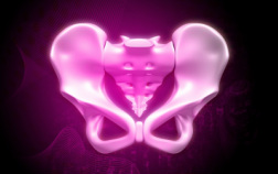 image: renjith krishnan
The one that has ruined my life! The one caused by an accident that was caused by EDS. The one that has put me in bad, unable to move, for five out of the last six year. Yes five, long years! Ironically, it's usually associated with athletes - I had to give up sport when I was 13.
See that little bit of cartilage where the pelvis meets in the front? That's the pubis symphysis where the problem is. For those of you who have given birth without epidural, imagine the pain of a head there. To the male population, imagine being kicked repeatedly in the groin. Consistently - every minute of every hour of every day for months and months on end. In my case, the pain spreads through my abdomen, which swells, and then it hurts to breathe. As the muscles move with each breath, it is agony. Doctors cannot tell me why this happens. If breathing hurts, then talking hurts too, so I become very isolated. The pain is made worse by sitting, standing, walking, sex, travelling by car, bus, rail and can also start for no apparent reason. Here's what Wikipedia says: "Since 1924, osteitis pubis has been known as a noninfectious inflammation of the pubis symphysis (also known as the pubic symphysis, symphysis pubis, or symphysis pubica) causing varying degrees of lower abdominal and pelvic pain. Osteitis pubis was first described in patients who had undergone suprapubic surgery and remains a well-known complication of invasive pelvis procedures . However, it may occur as an inflammatory process in athletes. The incidence and etiology of osteitis pubis as an inflammatory process versus an infectious process continues to fuel debate among physicians when confronted by a patient complaining of abdominal pain or pelvic pain and overlapping symptoms. Osteitis pubis may be diagnosed with an X-ray, where irregularity and widening of the pubic symphysis are hallmark findings. Similar change is also demonstrated with Computed Tomography (CT) however the multi-planar nature of CT has a higher sensitivity than X-rays. Though not well visualised on ultrasound, thickening of the superior joint capsule with cyst formation is a clue to the diagnosis, as well as secondary changes (tendinosis) of the adjacent adductor muscles (particularly adductor longus) and rectus abdominis. Ultrasound is also useful for excluding a hernia, which may co-exist with osteitis pubis and thus may also warrant additional treatment. Both ultrasound and CT may be used for injecting the pubic symphysis with corticosteroid as part of an athlete's treatment programme. Magnetic Resonance Imaging (MRI) combines the diagnostic advantages of CT and ultrasound, also showing bone marrow oedema and has the advantages of not being operator dependent, unlike ultrasound, nor does it use radiation, such as CT and X-rays. As such, MRI is the modality of choice." |
FOR MORE INFORMATION, HELP AND SUPPORT USE:
|
PELVIC INSTABILITY NETWORK SUPPORT
|
IF ANY CONTACTS ARE NO LONGER IN USE OR NEED UPDATING, OR IF YOU KNOW ANY MORE, GET IN TOUCH.
